Ultrasound Usage Beyond Pregnancy
As we all know, ultrasounds are used in pregnant women to determine fetal growth, development, and abnormalities, if any. Ultrasound imaging uses sound waves of high frequency to form an image of the fetal growth and the results of the test determine how you should work next. If the results are normal, then all good. And if it shows any abnormalities, you may need to seek further medical advice.
However, this diagnostic tool has several other applications. From diagnosing a wide variety of conditions with organs and soft tissues of the body to using it during medical procedures, ultrasounds have several uses. It is considered to be one of the safest way of imaging the internal organs of the body.
So, how does it work? What are its uses and types? Let’s take a look into it.
What is Ultrasound and How Does Ultrasound Imaging Work?
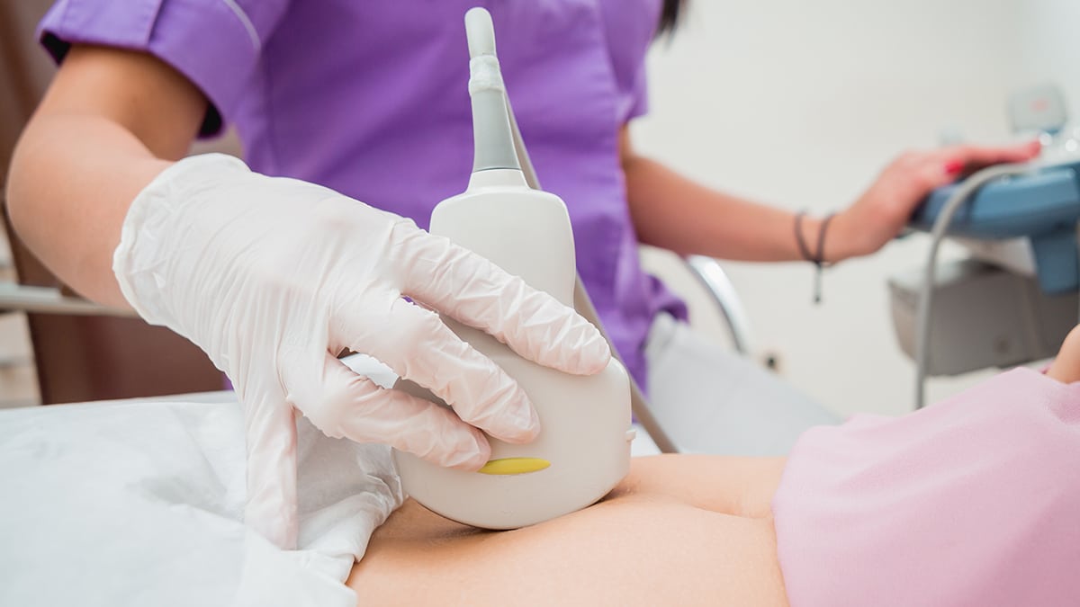
Ultrasounds are sound waves of high frequency. It is used in clinical process and diagnosis as it helps to form an image of the internal organs. The process uses an instrument called a transducer that’s placed directly on the skin or in some cases, inside a body opening. A thin layer of gel is applied to the skin to facilitate the transmission of the sound waves from the transducer into the body.
The transducer emits high-frequency sound waves and then records the echoes as the sound waves reflect back. In the process, it delivers the necessary information to produce an image of the soft tissues and organs on a computer screen.
The process is also known as sonography and ultrasound technician as a sonographer. The sonographer receives special training in performing the test. Once the test results are out, then that can be interpreted by a doctor and the necessary steps are taken.
Uses of Ultrasound Scans
As mentioned, ultrasound scans have a wide range of use. Doctors commonly use ultrasound scans to diagnose certain conditions in cases such as:
Pregnancy
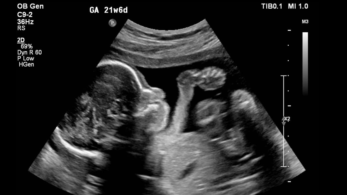
Ultrasounds have diverse uses during pregnancy. During the first trimester or the first three months, it can be used to check if the embryo is developing correctly. The test results will show if the embryo is developing inside the womb (and not inside a fallopian tube, for example). It also shows the number of embryos and is used to calculate the baby’s due date.
In the second trimester or tests performed between 18 and 20 weeks, development of fetal structures like limbs, brains can be found out. And the third trimester results show if the rate of growth of the baby is normal.
However, as the ultrasound images are not very clear, if some abnormalities are found in the test results, it must be followed and confirmed by some other tests. Results of sonography alone are not a clear determiner of a condition.
Diagnostics
Ultrasound imaging is used in diagnosing a wide variety of conditions with the soft tissues and organs in the body. Organs such as heart and blood vessels, liver, spleen, gallbladder, kidneys, uterus, ovaries, to name a few, are imaged using ultrasounds and looked for any abnormalities.
During Medical Procedures
Ultrasounds are often used in medical procedures such as needle biopsies where ultrasound is used to precisely remove tissue from inside the body. Ultrasound-guided needle placement (in blood vessels) is also common.
Types of Ultrasound
In most cases, ultrasound scans are done using a transducer on the skin surface. However, in some cases, doctors and technicians can choose to place a special transducer in one of the body’s natural openings for getting a clearer image.
- A transvaginal ultrasound better images of uterus and ovaries are obtained by placing a transducer in a woman’s vagina.
- In a transrectal ultrasound, a small lubricated probe is placed in the rectum for checking prostate conditions.
- In a transesophageal echocardiogram, a clearer image of the heart is obtained by placing the transducer probe in the esophagus.
In addition to what mentioned above, there are some other types of ultrasound imaging, such as:
- Doppler Ultrasound is used to visualize the blood flow through blood vessels, or other structures.
- Doppler fetal heart rate monitors are used to listen to the fetal heartbeat.
- Bone sonometry is used to assess bone fragility and possible osteoporosis.
- There is also a 3D imaging sonography. It uses technology to create three-dimensional images of organs rather than the traditional 2D image.
- In addition, there is also a 4D imaging ultrasound. It shows the 3D images in motion. This is particularly helpful for pregnant women to view their baby moving inside the womb.
Benefits of Ultrasound
Ultrasound scans have a lot of advantages:
- They do not require needles, injections, or incisions of any kind and are thus mostly painless.
- Ultrasound scans are much safer than diagnostic techniques like CT scans or X-rays. This is because, in an ultrasound scan, patients aren’t exposed to ionizing radiation, which is not the case with X-rays. In fact, no harmful effects of ultrasound are known when used by a certified sonographer or a doctor.
- Ultrasound imaging is less expensive than other methods.
Before an Ultrasound Scan
Depending on the type of ultrasound scan, your doctor may instruct you to not have any food or drinks several hours prior to the test. Or, to ensure that your bladder is full, you may be asked to drink several glasses of water and refrain from using the bathroom.
Procedure
You should wear comfortable clothing so that the technician can remove a part of it easily for placing the transducer. In some cases, you may need to wear a gown to facilitate the process.
The technician will apply a water-based gel on the area which is under scrutiny. And then glide the transducer over it for several minutes.
A typical ultrasound test lasts for about 30 minutes to an hour. You’ll be widely awake during the entire procedure and the procedure itself is generally not uncomfortable.
Immediately After the Scan
Immediately after the procedure, the technician will hand over paper towels or similar stuff with which you can rub off the gel. Next, you may get dressed and may need to use the bathroom.
Most ultrasound scans are non-invasive and thus there is no need for any recovery time. Generally, people can go about their way immediately after the scan is over.
Further Treatment
Ultrasound images, though are a good way of determining what’s going on inside the body, but it is not a clear determinator of any conditions. If some abnormalities are found in the scan results, it must be confirmed with further tests.
Final Thoughts
Ultrasound scan is a painless and quick method of imaging the internal organs and soft tissues. It has been around for years now and no after effects, harmful or not, are known. So, you can safely opt for this diagnosis method to find out any abnormalities with your internal organs.
All you need to do is connect with a reliable and certified treatment center offering ultrasound scans. So, if you are facing any symptoms and your doctor is prescribing you ultrasound scan, you should immediately do that without wasting any time. Delaying might complicate the situation or the symptoms might increase.
In any case, you should always consult with a medical professional first.
References:
https://www.betterhealth.vic.gov.au/health/conditionsandtreatments/ultrasound-scan
https://medlineplus.gov/lab-tests/sonogram/
https://www.fda.gov/radiation-emitting-products/medical-imaging/ultrasound-imaging
https://www.webmd.com/a-to-z-guides/what-is-an-ultrasound#2
https://www.betterhealth.vic.gov.au/health/healthyliving/pregnancy-tests-ultrasound