Select *DepartmentDoctor
Select Clinic *AestheticFacial Plastic SurgeryChiropracticDentistryDermatologyFamily MedicineGynecologyHijama & AcupunctureOrthopedicsPhysiotherapyPsychiatristSpeech & Language
Select Doctor *Grece El AsmarDr. Hakima AbdullahDr. SalmaDr. Iman FaresDr. Kate PanteleevaDr. Roaa TalalCraig ValentineDr. Sam KotkatDr. Angel MansoorDr. Lara Al-SammarraieDr. Heba AllabedDr. SumayaDr JulianaDr. Diyar MalikDr Aspasia MichalopoulouDr. JaniceDr. Mohamed Koutah
Preferred Appointment Date*
Select Time*MorningAfternoonEvening
I understand that this is not confirmed booking, will wait for confirmation message from the medical center
Why Annual Health Check-ups Matter: A Family Medicine Perspective
In the field of general medicine, the importance of annual health check-ups cannot be denied. Conducted by healthcare....
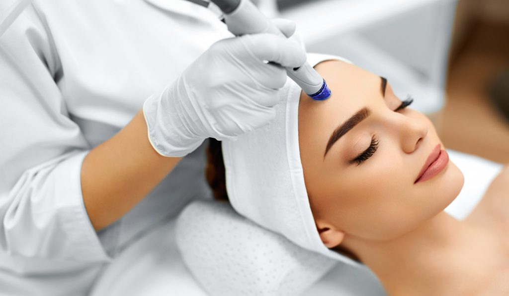
Endolift in Dubai | Non-Surgical Facelift at 7DMC, Dubai
Introduction: Redefining Skin Tightening Without Surgery Dubai’s aesthetic landscape is evolving rapidly, with more people seeking advanced, minimally....
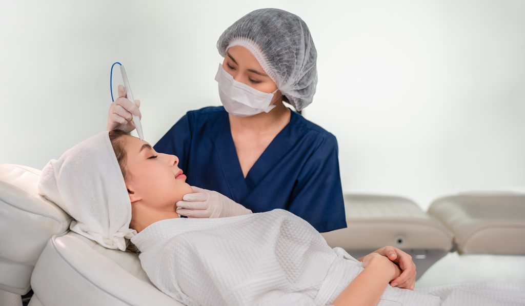
How to Properly Care for Skin after Cosmetic Procedures?
After months of research, you have selected the cosmetic procedure of your dreams, you have sorted an appointment....
Achieve Your Ideal Look with Best Aesthetic Clinic in Dubai
With Dubai being the pinnacle of modern developments and cutting-edge technology, it is no surprise that it has....
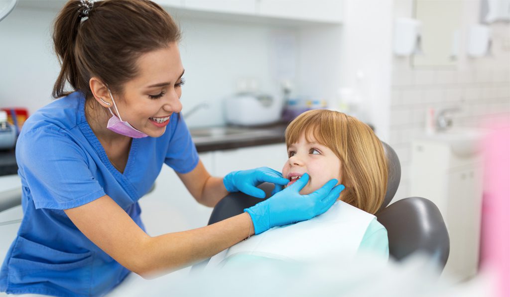
10 Signs that Your Child Needs to See a Dentist
Children go through massive transformative changes that need to be paid close attention to. Notable among them are....
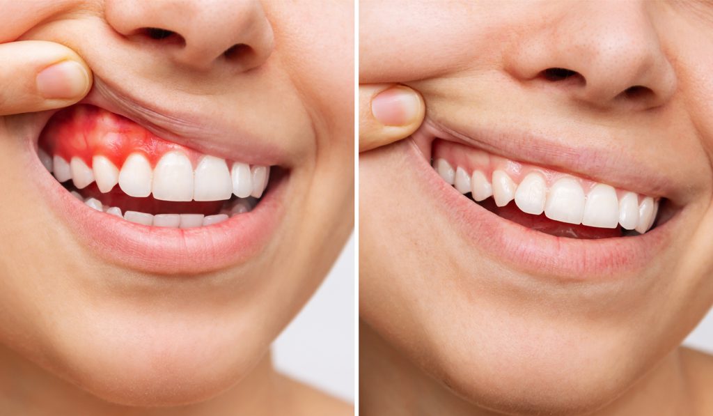
How to Overcome a Gummy Smile?
Your smile is one of the first indicators of positive body language and plays a crucial role in....
How to Choose Safe and Effective Aesthetic Treatments
Whether it is due to the influx of Dubai being a major fashion destination of the Middle East....
Top Questions to Ask Before Getting an Aesthetic Treatment
Being aware of your aesthetic needs and where you want to reach at the end of your aesthetic....
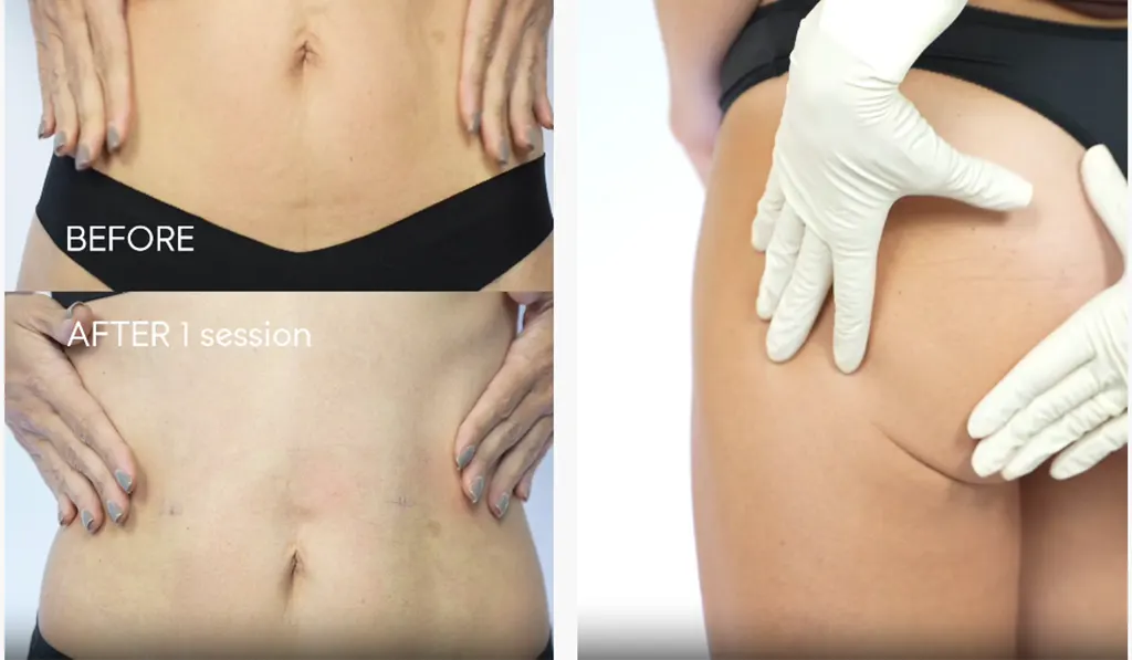
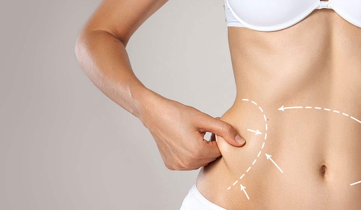
Is liposuction the best fat removal method?
Is liposuction the best fat removal method? Liposuction or lipoplasty is a surgical process where a suction procedure....
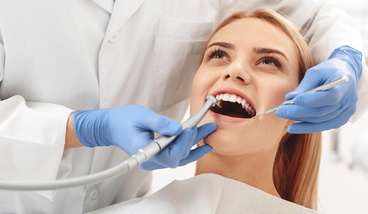
Why Dental Cleanings are Essential?
Dental Cleanings Your dentist will advise you that you need regular professional dental cleanings. Dental cleanings are not....
How to treat tailbone injury?
What is a tailbone injury and how to treat it? Tailbone discomfort can make routine activities painful at....
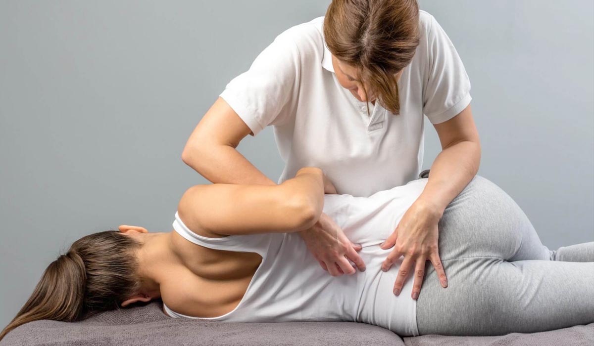
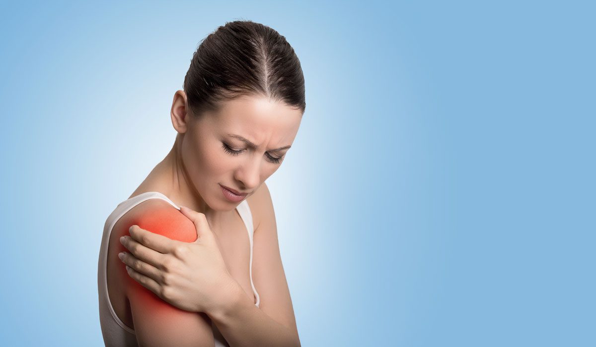
How to Deal with Shoulder Pain?
10 Ways to Deal with Shoulder Pain Shoulder pain is a common complaint that affects millions of people....
How to prevent premature aging?
How to prevent premature aging? Our bodies age naturally over time. Premature aging is when the signs of....
Are Dental X-Rays Safe During Pregnancy?
Are Dental X-Rays Safe During Pregnancy? Dental imaging is one major reason for anxiety among would-be parents and....
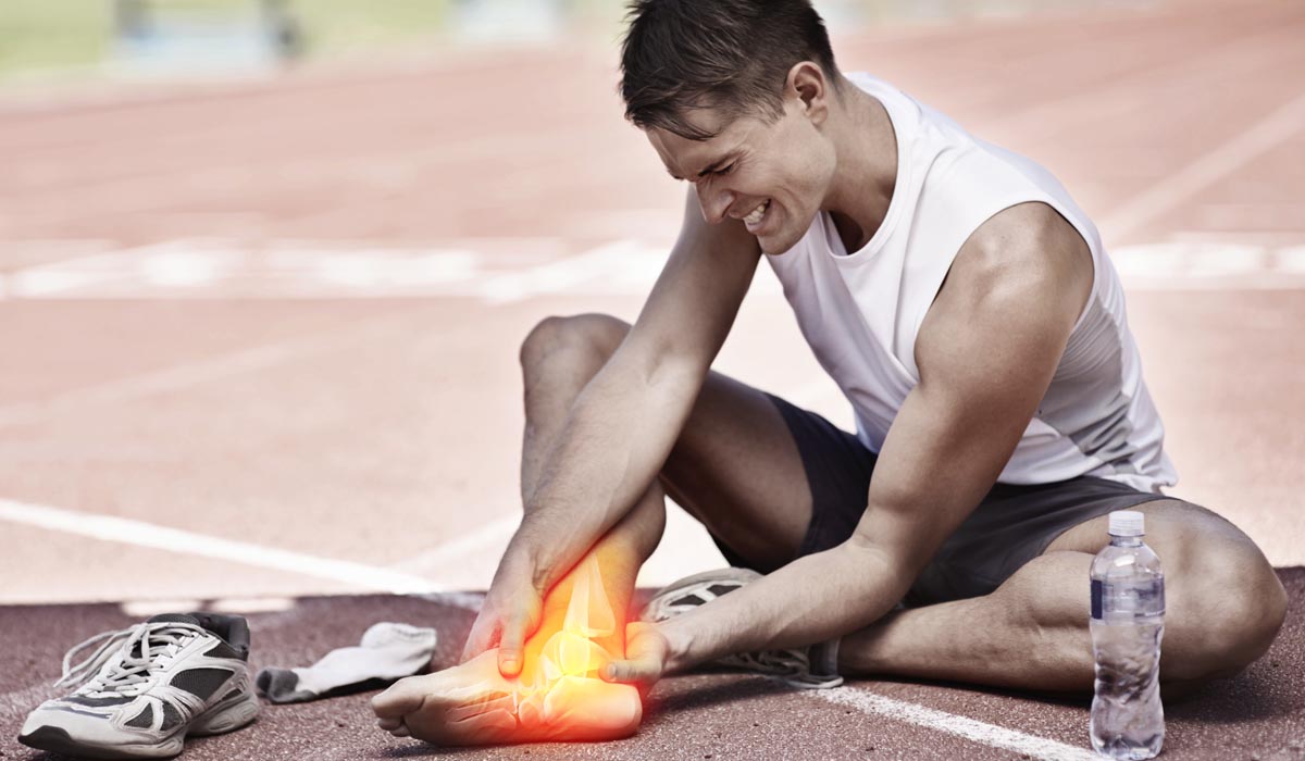
Common Sports Injuries and Ways to Prevent Them
10 Most Common Sports Injuries and Ways to Avoid Them Common sports injuries are typically brought on by....